Many veneer cases do not fail as a result of the laboratory’s work. Instead, they fail earlier during the planning, preparation, impressions, or putting together the details for the lab, so that, by the time the case reaches the lab, the problems are already present.
Veneers require careful preparation at every stage, and even seemingly small mistakes can lead to poor fit, weak bonding, unhappy patients, and costly remakes that could have been entirely avoided.
Successful veneer cases depend on clear communication, diligent preparation, and attention to detail from the very first step. When steps are missed or incorrect, even the most reputable of laboratories may not achieve the expected result.
Over-preparing the tooth
Over-preparing the tooth is one of the most common problems in veneer cases, as removing too much tooth structure can weaken the tooth and reduce the long-term success of the veneer. Additionally, veneers are designed to be conservative, so taking away more tooth than is needed can create problems before the case even reaches the laboratory.
When too much enamel is removed and goes deep into the dentine, bonding the veneers becomes less predictable compared to bonding to enamel. As well as increasing the risk of debonding, sensitivity, and the veneers failing over time, over-preparation can affect the final appearance of the veneers, making it harder to achieve a natural look and accurate shade match.
In some cases, where too much enamel has been removed, the laboratory will be limited in terms of options for creating the veneers. Technicians might need to make a thicker veneer to compensate for the space created, which in turn can affect the final appearance and fit. In many cases, preserving as much healthy enamel as possible gives both the dentist and technician a better foundation for a predictable, long-lasting result.
Incomplete bite scans
Accurate bite scans are essential so that dental labs can produce veneers that fit properly and function comfortably. When scans are incomplete or wrong, a dental laboratory might struggle to understand important elements like the patient’s occlusion and how the restorations should sit against the opposing teeth.
A frequent issue dental laboratories encounter is unclear preparation margins. If the margin is not fully visible in the scan, the technician may find it hard to design a restoration with the level of precision they would like, resulting in poor fit, open margins, or adjustments needing to be made at the fitting appointment. Poor soft tissue retraction is often a major cause of this problem, especially when the gingiva covers part of the preparation area, or moisture affects the scan quality.
Incomplete bite registrations can also create problems with contact points and occlusion. If the scan does not accurately capture how the patient bites together, the final veneers might feel too high or uncomfortable when fitted, so careful scanning, good moisture control, and retraction all help make sure the lab gets the information it needs to produce a good result for the patient.
Lack of stump shade photos
Stump shade photos play an important role in veneer cases, especially because veneers are most commonly sought for cosmetic purposes to improve the appearance of a person’s smile. Without clear information, a dental laboratory may struggle to predict how the final veneers will appear once they have been bonded to the prepared teeth.
Because the colour of the underlying tooth can strongly influence the final result, it is crucial that the laboratory receives clear stump shade photos. Without these, the technician has less information to work with when planning the ceramic build-up, which can be a problem in cases involving dark teeth, previous restorations, or uneven discolouration. Even when a particular veneer shade is chosen, the final result can still look different in the mouth.
Thin veneers in particular are affected by the colour underneath because ceramic materials still allow some light to pass through. Without accurate stump shade records, achieving a natural and balanced appearance becomes more difficult, so good-quality photos taken under consistent lighting conditions help the laboratory create the best possible veneers.
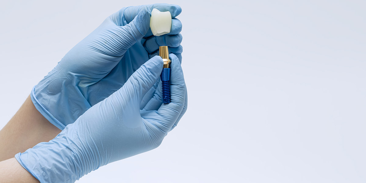
Wrong choice of material
The material used for veneers can affect both how they look and perform. Different materials behave in different ways, so the material needs to suit the specific case. Some are more translucent and natural-looking, while others are better at masking dark or discoloured teeth, but choosing the wrong option can make the veneer look too opaque, too grey, or unnatural.
Strength is also an important factor, as certain materials work well for thin veneers and conservative preparations, while others are more suitable for patients with heavier bites or less enamel available for bonding.
The most common materials used to make veneers are:
| Material | Thickness | Strength | Appearance | Stain resistance | Lifespan |
| Porcelain | 0.5 to 0.7mm | High | Excellent | Very high | 10 to 20 years (and beyond) |
| Lithium discilicate | 0.3 to 0.7mm | Very high | First-rate | Very high | 10 to 20 years (and beyond) |
| Composite | 0.3 to 0.5mm | Moderate | Good | Moderate | 5 to 7 years |
| Ultra-thin aesthetic ceramics | 0.2 to 0.5mm | Moderate | Excellent | High | 7 to 15 years |
If the material chosen is not a good fit for a patient’s needs, problems like chipping, poor aesthetics, or early failure become more likely, so careful planning and collaboration between the dentist and laboratory help avoid this.
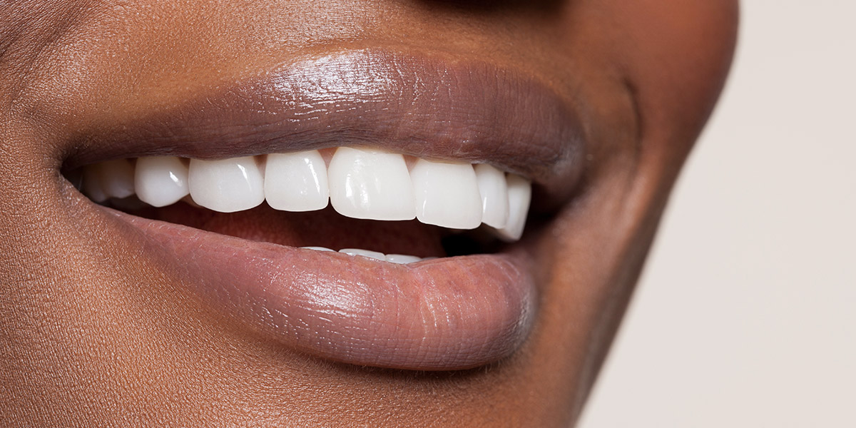
Unrealistic patient expectations
Unrealistic expectations can cause problems in veneer cases long before any work starts, often developing from what patients see on social media. Platforms like Instagram and TikTok are full of transformation videos and images of perfectly straight smiles that are often filtered, edited, and taken in ideal lighting and from carefully chosen angles. These can lead patients to believe that they, too, could achieve these results from veneers.
In reality, however, every smile is different, and factors like tooth colour, bite, gum levels, and the position of natural teeth can all affect what veneers can realistically achieve. A result that looked simple online may actually demand complex planning, or may not be suitable for everyone.
When expectations are based on filtered or idealised content, there is a higher risk of disappointment even if the clinical work is successful. A technically good outcome can still feel underwhelming if it doesn’t match what the patient saw online.
Clear communication from the start helps bridge this gap. Showing real-life case examples and explaining limitations in a simple, honest way helps patients understand what is achievable and leads to more realistic expectations.
Creating a pre-submission checklist
A clear pre-submission checklist helps reduce errors before a veneer case is sent to the dental laboratory, as it ensures all the key clinical information is complete and ready for the technician who picks up the case.
A good checklist might include:
- Full arch scans with clear detail
- Accurate bite registration showing a patient’s occlusion
- Clear preparation margins
- Good soft tissue retraction to expose the full prep
- Pre-operative photos for reference
- Stump shade photographs taken after preparation
- Final shade selection
- Close-up photos with shade tabs in place
- Clear prescription form completed in full, with notes on any special requests or concerns
When important details are missing, the laboratory may have to ask for more information, which can delay the case. Using the same checklist each time helps both parties understand what to expect, as well as what is expected of them, and improves overall case quality.
Give your patients the best veneers possible
Good veneer outcomes rely on everything working together clearly and consistently from the initial planning right through to the final fitting, and beyond. When each step is done properly, it becomes much easier to achieve results that fit well, look natural, and feel comfortable for the patient.
Small details in communication and preparation can make a noticeable difference to the process. When the dental team and our team at GoDigital Dental are working with the right information at the right time, it reduces problems and keeps cases on track.
Ultimately, better organisation and attention to detail throughout the process lead to more predictable results and benefit both the clinician and the patient, with fewer surprises and a more positive overall experience.
FAQs
How much tooth should be removed for veneers?
Tooth preparation for veneers is usually kept very light, as the idea is to be as conservative as possible while still achieving the planned result. In most cases, only a thin layer of enamel, around 0.3 to 0.5 mm, is removed. This is just enough to create space for the veneer while keeping the tooth as natural as possible.
There are sometimes situations where a little more reduction is needed, usually if the tooth is very dark or if a patient wants a more dramatic change in the shape or position of their new teeth. Even where more enamel removal is needed, the goal is always to avoid removing more tooth than necessary, as preserving enamel helps the veneer bond more securely and keeps the tooth stronger in the long term.
What is the best way to get good gum retraction for scans?
To achieve good gum retraction for scans, it is important to gently move the gum tissue away from the tooth before scanning. This can be done using retraction cord, retraction paste, or a combination of both, depending on the case. The goal is to clearly expose the full preparation and margins.
Good moisture control is also essential, as saliva or bleeding can affect scan accuracy, and the tissue should also be stable and dry enough to allow the dentist to capture a clear image without distortion. Careful handling of the gums avoids trauma, helping maintain a clean field and improving the quality of the final scan data for the laboratory.
Which details need to be included in a prescription form?
A prescription form for veneer cases should include all key clinical and technical details needed by the laboratory, including patient information, tooth numbers, the shade they have chosen, and the type of restoration required. It should also clearly state the material choice, any special instructions, and the desired final outcome for each tooth.
Make sure to include notes on preparation design, bite considerations, and any specific aesthetic requirements. Supporting information, such as photographs, scans, and stump shade details, should be referenced and sent along as specified by the lab.
A complete prescription and clear supporting documentation help reduce errors, avoid delays, and give the laboratory everything they need to produce consistent, accurate restorations that match the clinical plan and meet patients’ expectations.