
Restoring a Compromised Tooth Through Digital Dentistry and Clinical Collaboration
A restorative case by Dr Lewis Dearsley of Sync Dental Windsor, in collaboration with GoDigital Dental Laboratory, Nottingham
Case Summary
Case: Restoration of a fractured upper right second premolar (UR5)
Treatment: Deep margin elevation, root canal treatment, composite core reconstruction and cuspal coverage
Workflow: Pre-operative and preparation scans, digital CAD design, CAD/CAM milling and ceramic finishing
Restoration: IPS e.max vonlay
Outcome: Preservation and restoration of a structurally compromised natural tooth
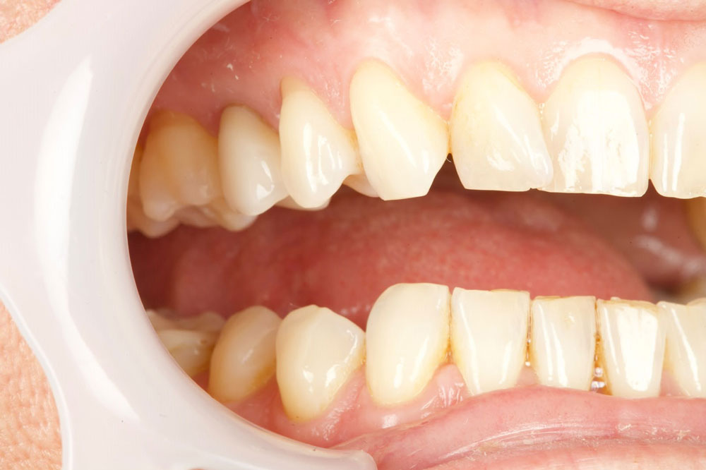
Preserving a Natural Tooth Following a Significant Fracture
When a tooth fractures below the gumline, the first challenge is deciding whether it can be restored. Much depends on the extent of the fracture, how accessible the margin is and how much healthy tooth structure remains.
In this case, Dr Lewis Dearsley of Sync Dental Windsor treated a patient with a fractured palatal cusp affecting the upper right second premolar (UR5). The fracture extended subgingivally and involved pulpal exposure, raising concerns about the tooth’s restorability.
Although the extent of the fracture initially raised doubts about whether the tooth could be saved, Dr Dearsley was able to manage the deep margin and proceed with treatment.
GoDigital Dental Laboratory worked with Dr Dearsley to produce an IPS e.max vonlay for the prepared tooth. Using the same CAD/CAM technology and ceramic techniques involved in producing dental crowns and bridges, the team designed, milled and finished the restoration in-house.
Assessing the Fracture and Establishing Restorability
The patient presented with a substantial fracture of the palatal cusp, extending beneath the gingival margin and exposing the dental pulp.
With the fracture extending below the gumline, access to the palatal margin was difficult. Before committing to further treatment, Dr Dearsley needed to establish whether the tooth was restorable.
A gingivectomy was carried out before rubber dam placement to improve access. Deep margin elevation (DME) was then used to raise the palatal margin to a more accessible position.
Once the deep margin had been raised, the tooth was considered restorable and Dr Dearsley could proceed with treatment.
Initial Fractured Premolar

Pre-Treatment Clinical View

Root Canal Treatment and Composite Core Reconstruction
Once restorability had been established, root canal treatment was completed before the definitive restoration.
The compromised tooth structure was then rebuilt using a composite core and dentine replacement, incorporating everX Posterior and Kuraray AP-X.
This reconstruction provided the foundation for the final indirect restoration.
UR5 was then prepared for an IPS e.max vonlay to provide cuspal coverage and protect the remaining tooth structure.
Following preparation, digital scans were supplied to GoDigital Dental Laboratory to begin the design and manufacturing stages.
Digital Preparation Scan

Prepared Tooth Scan

Digital Scanning and CAD Design
Dr Dearsley supplied GoDigital with two digital scans. The first was taken at the time of whitening and showed the pre-operative tooth anatomy, including the large palatal fracture. The second was taken once UR5 had been prepared for the vonlay.
Using these digital dental impressions, Moritz Pohle, GoDigital’s CAD/CAM Technician, overlaid the pre-operative and preparation scans to compare the tooth before and after preparation.
By comparing the two scans, Moritz was able to develop a patient-specific CAD design that accounted for the remaining tooth structure, required cuspal coverage and relationship with the surrounding dentition.
The final CAD design established the shape of the vonlay, replacing the fractured tooth structure and fitting around the remaining natural tooth.
Once the design was complete, the case moved on to milling and ceramic finishing.
Digital Vonlay CAD Design


IPS e.max Milling and Restoration Manufacture
Following the digital design stage, the restoration was milled from IPS e.max, a lithium disilicate glass-ceramic widely used for indirect dental restorations.
IPS e.max offers a combination of strength and aesthetic properties, making it a suitable restorative material for a range of ceramic crowns, onlays and other indirect restorations.
The vonlay was milled to match the CAD design and provide the cuspal coverage required for UR5. Once milled, it was passed to the ceramic department for finishing.
The CAD/CAM workflow enabled the laboratory to translate the digital design into a patient-specific ceramic restoration, ready for the final finishing stages.
IPS e.max Vonlay at Fitting

What Is an IPS e.max Vonlay?
An IPS e.max vonlay is a type of partial ceramic restoration that combines elements of a veneer and an onlay. It covers the biting surface and one or more cusps of a tooth, extending onto the outer surface where required.
Made from lithium disilicate glass-ceramic, IPS e.max combines strength with a natural, tooth-coloured appearance. It is commonly used for indirect restorations where both durability and aesthetics are important.
The main characteristics of an IPS e.max vonlay include:
- Preservation of natural tooth structure: Unlike a full-coverage crown, a vonlay can restore damaged areas without covering the entire tooth, allowing more healthy tooth structure to be retained.
- Strength and durability: Lithium disilicate is a strong ceramic material suitable for restoring teeth that need to withstand everyday biting and chewing forces.
- Natural appearance: The ceramic can be shade-matched to the surrounding teeth, with translucency that helps the restoration blend with natural enamel.
- Adhesive bonding: The restoration is bonded to the prepared tooth using dental adhesive techniques, helping to provide a secure fit.
When is a vonlay used?
Vonlays may be used to restore fractured, cracked or heavily worn teeth, or to replace large, failing restorations. They are particularly useful when both the biting surface and an outer surface of the tooth need to be restored.
Each vonlay is individually designed to fit the prepared tooth, taking into account the remaining tooth structure and the patient’s bite.
Ceramic Finishing and Quality Control
Once the IPS e.max vonlay had been milled, it was ready for the final stages of ceramic finishing and quality control at GoDigital Dental Laboratory.
The restoration was finished by Tilly, a second-year Dental Technician Apprentice, who prepared the ceramic vonlay for its final checks.
Jenna Ellis, Head of Ceramics, then carried out the quality control checks before the completed restoration was sent to Dr Dearsley at Sync Dental Windsor for the patient’s fitting appointment.

Fitting the IPS e.max Vonlay
Following completion of the clinical preparation and laboratory manufacturing stages, Dr Dearsley fitted the finished IPS e.max vonlay.
The finished vonlay provided cuspal coverage for UR5, restoring the tooth following the earlier deep margin elevation, root canal treatment and composite reconstruction.
Clinical photographs taken immediately after fitting document the completed restoration.
Further photographs taken two weeks after fitting show its appearance alongside the surrounding natural teeth.
Despite the extent of the original fracture, the natural tooth was retained and restored. Both Dr Dearsley and the patient were delighted with the result.
Preserving Natural Tooth Structure Through Digital Dentistry

This case shows how careful assessment and management of a deep fracture can make it possible to restore a tooth that initially appears difficult to save.
Dr Dearsley’s clinical work established a restorable tooth, while GoDigital Dental Laboratory used the pre-operative and preparation scans to design and manufacture the IPS e.max vonlay.
The result was a ceramic restoration providing cuspal coverage while retaining the patient’s natural premolar. It’s also a good example of how close communication between dentist and laboratory can help with more challenging restorative cases.
Case Study Before
Case Study Design

Case Study After

“None of this would have been possible without the fantastic work of the team at GoDigital Dental Laboratory. Their skill and attention to detail made all the difference, the patient and I are absolutely delighted with the final result.”
Dr Lewis Dearsley
Associate Dentist
Sync Dental Windsor
Credits to:
Digital CAD design:
Moritz Pohle, CAD/CAM Technician
Ceramic finishing:
Tilly, Dental Technician Apprentice
Quality control: Jenna Ellis, Head of Ceramics
Our Dental Lab Services
Advanced dental implant-based solutions for single implant, multiple implants, full-arch, All-on-X, full mouth restorations.
Crowns and bridges manufactured from monolithic zirconia, lithium disilicate, feldspar and hybrid ceramics.
Expertly crafted dental veneers, using advanced digital workflows, high strength premium materials, and precise CAD/CAM technology.
A professional implant planning service designed to give clinicians greater confidence, accuracy and predictability.
A bespoke shade taking service at our dental lab in Nottingham, with experienced technicians creating accurate dental products.
Using advanced technology we create a virtual image that allows both the dentist and patient to see the results before treatment begins.